In a letter to the editor, asking how many nerves supply the shoulder, Dr Price stated “… any residual pain in postanesthesia care unit tends to be felt anteriorly. This usually settles after 1 to 2 hours and seems to correlate with resolution of the arthroscopic fluid–induced distension of the shoulder capsule and thus be mediated by anterior articular branches. But which ones?”
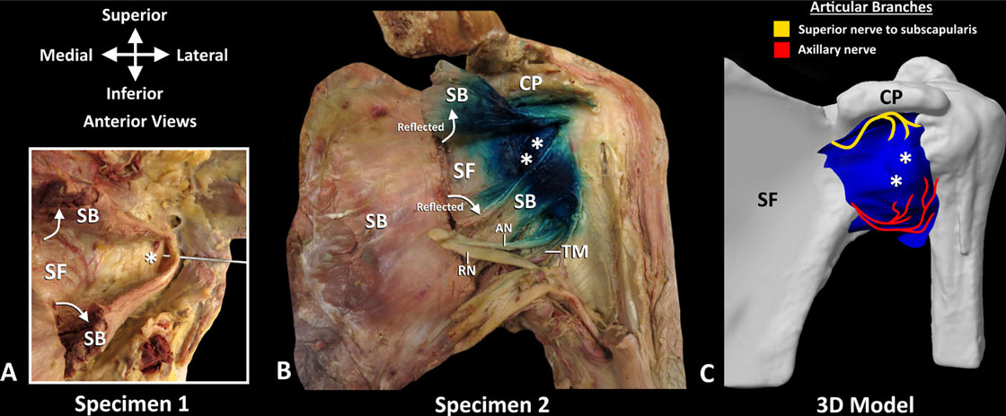
In light of the findings of our recent article, investigating the innervation of the glenohumeral joint (GHJ) capsule, we found that on reflection of subscapularis the anterior capsule was innervated by articular branches of the superior nerve to subscapularis and axillary nerve. To further investigate the possibility of dye spread to capture these articular branches deep to subscapularis, as a proof of concept, two lightly embalmed cadaveric shoulders were needled using ultrasound (US) guidance. In the first specimen, one needle was placed to determine if it is possible to target the plane between the subscapularis and the anterior capsule overlying the anterior rim of the glenoid fossa. Following placement of the needle, the specimen was dissected and the subscapularis reflected. The needle was found in the plane between subscapularis
and anterior GHJ capsule (figure 1A). This observation led us to attempt injecting methylene blue (2 mL) using US guidance into this plane, in a second specimen, to
determine if the articular branches could be captured. On dissection, the dye spread
into this plane stained the entire anterior joint capsule supplied by articular branches of the superior nerve to subscapularis and axillary nerve (figure 1B,C). In addition, the main trunk of axillary nerve was also stained. These anatomical findings need
further clinical investigation.
Although we have never had the pleasure of meeting Dr Darcy Price, we have read with great interest his insightful letters regarding shoulder analgesia. As a tribute to Dr Price’s contribution and efforts towards optimizing postoperative shoulder analgesia, we would like to dedicate the Deep Plane to Subscapularis analgesia approach as the Darcy Price Salutation block.
John Tran, Anne Agur, Philip Peng
Division of Anatomy, Department of Surgery, University of Toronto, Toronto, Ontario, Canada Department of Anesthesia, Toronto Western Hospital, University Health Network, Toronto, Ontario, Canada
Correspondence to John Tran, Division of Anatomy, Department of Surgery, University of Toronto, Toronto, ON M5S 1A8, Canada; johnjt.tran@mail.utoronto.ca
Contributors JT, AA, and PP contributed equally to this letter.
Funding The authors have not declared a specific grant for this research from any funding agency in the public, commercial or not-for-profit sectors.
Competing interests None declared. Patient consent for publication Not required. Provenance and peer review Not commissioned; internally peer reviewed.
© American Society of Regional Anesthesia & Pain Medicine 2019. No commercial re-use. See rights and permissions. Published by BMJ.

Figure 1 Dye spread in deep plane to subscapularis (A) Dissection of deep plane to subscapularis following needle placement. Note the subscapularis was cut and reflected superiorly and inferiorly to expose the anterior glenohumeral joint capsule (*). (B) Dissection following dye injection using the deep plane to subscapularis approach. The dye was seen spreading in the deep subscapular plane over the glenohumeral joint capsule (**). Note in this picture, only 2 mL of methylene blue was injected, and dye was seen staining the main trunk of the axillary nerve. (C) 3D modeling of dye spread. Asterisk (white) indicates anterior glenohumeral joint capsule. 3D, three-dimensional; AN, axillary nerve; CP, coracoid process; SB, subscapularis; SF, subscapular fossa. RN, radial nerve; TM, teres major tendon.
To cite Tran J, Agur A, Peng P. Reg Anesth Pain Med Epub ahead of print: [please include Day Month Year]. doi:10.1136/rapm-2019-100496
Received 23 February 2019
Accepted 8 March 2019
Reg Anesth Pain Med 2019;0:1.
doi:10.1136/rapm-2019-100496
1 Price D. How many nerves supply the shoulder? Reg Anesth Pain Med 2018;43.
2 Tran J, Peng PWH, Agur AMR. Anatomical study of the innervation of glenohumeral and acromioclavicular joint capsules: implications for image-guided intervention. Reg Anesth Pain Med 2019.
3 Price D. Optimizing the combined suprascapular and axillary nerve (SSAX) block. Reg Anesth Pain Med 2017;42.
4 Price DJ. Toward a better understanding of brachial plexus anatomy for shoulder, Forearm, and hand anesthesia. Reg Anesth Pain Med 2013;38.
5 Price DJ. Combined Suprascapular and axillary nerve blockade. Arthroscopy 2008;24:1316–7.
